Figure 1
Concentration Polarization of Ox-LDL and Its Effect on Cell Proliferation and Apoptosis in Human Endothelial Cells
Shijie Liu*, Jawahar L Mehta, Yubo Fan, Xiaoyan Deng and Zufeng Ding*
Published: 30 December, 2016 | Volume 1 - Issue 1 | Pages:

Figure 1:
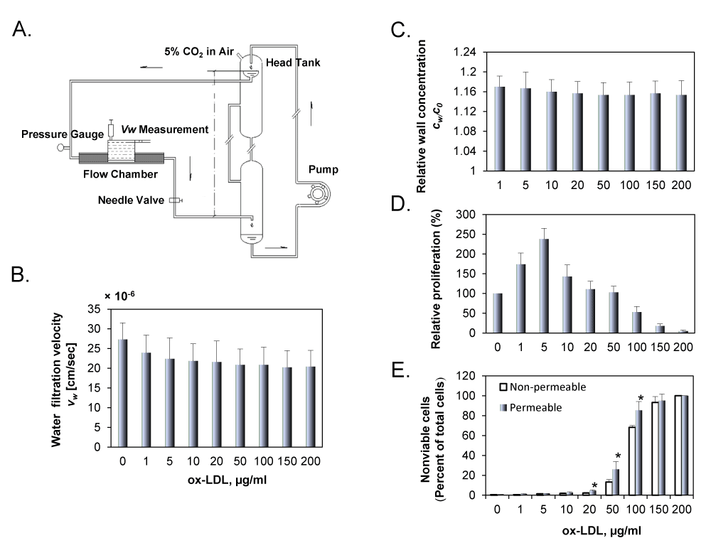
A. Schematic drawing of the perfusion system. The overflow head-tank provided a steady flow to the parallel-plate flow chamber. The filtration rate measurement cell on the abluminal side of the culture was filled with the same fluid as the perfusate. B. Water filtration rate for the permeable group with concentration of DiIox-LDL varied from 1 to 200 μg/ml. C. Wall concentration of DiI-ox-LDL in permeable group was assessed by measuring the fluorescence intensity of DiI-ox-LDL using a confocal laser microscope. All data were normalized with the bulk concentration, c0. D. Effect of ox-LDL on EC proliferation in endothelial cells was assessed by MTT assay. E. Trypan blue staining data in ECs cultured on permeable or non-permeable membranes and treated with ox-LDL (1 to 200 μg/ml). EC suspensions were exposed to perfusion pressure of 100 mmHg for 24 h. The results are expressed as means ± SD (n= 6). *P< 0.05 between permeable and non-permeable groups.
Read Full Article HTML DOI: 10.29328/journal.jccm.1001003 Cite this Article Read Full Article PDF
More Images
Similar Articles
-
Concentration Polarization of Ox-LDL and Its Effect on Cell Proliferation and Apoptosis in Human Endothelial CellsShijie Liu*,Jawahar L Mehta,Yubo Fan,Xiaoyan Deng,Zufeng Ding*. Concentration Polarization of Ox-LDL and Its Effect on Cell Proliferation and Apoptosis in Human Endothelial Cells. . 2016 doi: 10.29328/journal.jccm.1001003; 1:
-
Indications and Results of Coronarography in Senegalese Diabetic Patients: About 45 CasesNdao SCT*,Gaye ND,Dioum M,Ngaide AA,Mingou JS,Ndiaye MB, Diao M,Ba SA. Indications and Results of Coronarography in Senegalese Diabetic Patients: About 45 Cases. . 2017 doi: 10.29328/journal.jccm.1001007; 2: 013-019
-
Spontaneous rupture of a giant Coronary Artery Aneurysm after acute Myocardial InfarctionOğuzhan Çelik,Mucahit Yetim,Tolga Doğan,Lütfü Bekar,Macit Kalçık*,Yusuf Karavelioğlu. Spontaneous rupture of a giant Coronary Artery Aneurysm after acute Myocardial Infarction. . 2017 doi: 10.29328/journal.jccm.1001009; 2: 026-028
-
Investigation of Retinal Microvascular Findings in patients with Coronary Artery DiseaseTolga Doğan*,Osman Akın Serdar,Naile Bolca Topal,Özgür Yalçınbayır. Investigation of Retinal Microvascular Findings in patients with Coronary Artery Disease. . 2017 doi: 10.29328/journal.jccm.1001012; 2: 042-049
-
Lipid-induced cardiovascular diseasesSumeet Manandhar,Sujin Ju,Dong-Hyun Choi,Heesang Song*. Lipid-induced cardiovascular diseases. . 2017 doi: 10.29328/journal.jccm.1001018; 2: 085-094
-
Glycosaminoglycans as Novel Targets for in vivo Contrast-Enhanced Magnetic Resonance Imaging of AtherosclerosisYavuz O Uca*,Matthias Taupitz. Glycosaminoglycans as Novel Targets for in vivo Contrast-Enhanced Magnetic Resonance Imaging of Atherosclerosis. . 2020 doi: 10.29328/journal.jccm.1001091; 5: 080-088
-
Correlation between chronic inflammation of rheumatoid arthritis and coronary lesions: “About a monocentric series of 202 cases”Nassime Zaoui*,Amina Boukabous,Nabil Irid,Nadhir Bachir,Ali Terki. Correlation between chronic inflammation of rheumatoid arthritis and coronary lesions: “About a monocentric series of 202 cases”. . 2022 doi: 10.29328/journal.jccm.1001144; 7: 109-114
Recently Viewed
-
Are Biofungicides a Means of Plant Protection for the Future?Radek Vavera*, Josef Hýsek. Are Biofungicides a Means of Plant Protection for the Future?. J Plant Sci Phytopathol. 2024: doi: 10.29328/journal.jpsp.1001130; 9: 041-042
-
A Mini Review of Newly Identified Omicron SublineagesDasaradharami Reddy K*,Anusha S,Palem Chandrakala. A Mini Review of Newly Identified Omicron Sublineages. Arch Case Rep. 2023: doi: 10.29328/journal.acr.1001082; 7: 066-076
-
New Fungi Associated with Blackberry Root Rot (Rubus spp.), in Michoacán, MexicoLuis Mario Tapias Vargas, Anselmo Hernández Pérez, Adelaida Stephany Hernández Valencia*. New Fungi Associated with Blackberry Root Rot (Rubus spp.), in Michoacán, Mexico. J Plant Sci Phytopathol. 2024: doi: 10.29328/journal.jpsp.1001129; 8: 038-040
-
Haemodynamic, Biochemical and Respiratory Implications of total Bronchoalveolar Lavage in Pulmonary Alveolar ProteinosisMaría Nieves Balaguer Cartagena*, Ester Villareal Tello, Begoña Balerdi Pérez, Andrés Briones Gómez, Raquel Martínez Tomás, Enrique Cases Viedma. Haemodynamic, Biochemical and Respiratory Implications of total Bronchoalveolar Lavage in Pulmonary Alveolar Proteinosis. Arch Case Rep. 2023: doi: 10.29328/journal.acr.1001071; 7: 023-028
-
Towards A 21st Century Systematize the Ideas; COVID-19, Sustainability and Discourse of SDG, (Sustainable Development Goals), The Cities and Housing ModelsHülya Coskun*. Towards A 21st Century Systematize the Ideas; COVID-19, Sustainability and Discourse of SDG, (Sustainable Development Goals), The Cities and Housing Models. Arch Case Rep. 2024: doi: 10.29328/journal.acr.1001089; 8: 027-035
Most Viewed
-
Evaluation of Biostimulants Based on Recovered Protein Hydrolysates from Animal By-products as Plant Growth EnhancersH Pérez-Aguilar*, M Lacruz-Asaro, F Arán-Ais. Evaluation of Biostimulants Based on Recovered Protein Hydrolysates from Animal By-products as Plant Growth Enhancers. J Plant Sci Phytopathol. 2023 doi: 10.29328/journal.jpsp.1001104; 7: 042-047
-
Feasibility study of magnetic sensing for detecting single-neuron action potentialsDenis Tonini,Kai Wu,Renata Saha,Jian-Ping Wang*. Feasibility study of magnetic sensing for detecting single-neuron action potentials. Ann Biomed Sci Eng. 2022 doi: 10.29328/journal.abse.1001018; 6: 019-029
-
Physical activity can change the physiological and psychological circumstances during COVID-19 pandemic: A narrative reviewKhashayar Maroufi*. Physical activity can change the physiological and psychological circumstances during COVID-19 pandemic: A narrative review. J Sports Med Ther. 2021 doi: 10.29328/journal.jsmt.1001051; 6: 001-007
-
Pediatric Dysgerminoma: Unveiling a Rare Ovarian TumorFaten Limaiem*, Khalil Saffar, Ahmed Halouani. Pediatric Dysgerminoma: Unveiling a Rare Ovarian Tumor. Arch Case Rep. 2024 doi: 10.29328/journal.acr.1001087; 8: 010-013
-
Prospective Coronavirus Liver Effects: Available KnowledgeAvishek Mandal*. Prospective Coronavirus Liver Effects: Available Knowledge. Ann Clin Gastroenterol Hepatol. 2023 doi: 10.29328/journal.acgh.1001039; 7: 001-010

HSPI: We're glad you're here. Please click "create a new Query" if you are a new visitor to our website and need further information from us.
If you are already a member of our network and need to keep track of any developments regarding a question you have already submitted, click "take me to my Query."


