Figure 3
Pathological left ventricular hypertrophy and outflow tract obstruction in an infant of a diabetic mother: A case report
Ujuanbi AS*, Onyeka CA, Yeibake WS, Oremodu T, Kunle-Olowu OE and Otaigbe BE
Published: 03 March, 2020 | Volume 5 - Issue 1 | Pages: 047-050
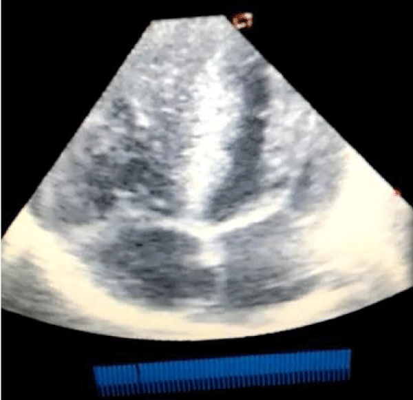
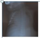
Figure 3:
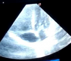
Echocardiography (apical 4 chamber view) showing severe biventricular and septal hypertrophy causing left ventricular outflow tract obstruction and a markedly diminished left ventricular cavity.
Read Full Article HTML DOI: 10.29328/journal.jccm.1001085 Cite this Article Read Full Article PDF
More Images
Similar Articles
-
Pathological left ventricular hypertrophy and outflow tract obstruction in an infant of a diabetic mother: A case reportUjuanbi AS*,Onyeka CA,Yeibake WS, Oremodu T,Kunle-Olowu OE,Otaigbe BE. Pathological left ventricular hypertrophy and outflow tract obstruction in an infant of a diabetic mother: A case report. . 2020 doi: 10.29328/journal.jccm.1001085; 5: 047-050
Recently Viewed
-
Exceptional cancer responders: A zone-to-goDaniel Gandia,Cecilia Suárez*. Exceptional cancer responders: A zone-to-go. Arch Cancer Sci Ther. 2023: doi: 10.29328/journal.acst.1001033; 7: 001-002
-
Knowledge, Attitude, and Practice of Healthcare Workers in Ekiti State, Nigeria on Prevention of Cervical CancerAde-Ojo Idowu Pius*, Okunola Temitope Omoladun, Olaogun Dominic Oluwole. Knowledge, Attitude, and Practice of Healthcare Workers in Ekiti State, Nigeria on Prevention of Cervical Cancer. Arch Cancer Sci Ther. 2024: doi: 10.29328/journal.acst.1001038; 8: 001-006
-
Update on Mesenchymal Stem CellsKhalid Ahmed Al-Anazi*. Update on Mesenchymal Stem Cells. J Stem Cell Ther Transplant. 2024: doi: 10.29328/journal.jsctt.1001035; 8: 001-003
-
Impact of Chronic Kidney Disease on Major Adverse Cardiac Events in Patients with Acute Myocardial Infarction: A Retrospective Cohort StudyAbbas Andishmand, Ehsan Zolfeqari*, Mahdiah Sadat Namayandah, Hossein Montazer Ghaem. Impact of Chronic Kidney Disease on Major Adverse Cardiac Events in Patients with Acute Myocardial Infarction: A Retrospective Cohort Study. J Cardiol Cardiovasc Med. 2024: doi: 10.29328/journal.jccm.1001175; 9: 029-034
-
Myxedema Coma and Acute Respiratory Failure in a Young Child: A Case ReportRinah Elaisse R Dolores, Marion O Sanchez*. Myxedema Coma and Acute Respiratory Failure in a Young Child: A Case Report. Ann Clin Endocrinol Metabol. 2023: doi: 10.29328/journal.acem.1001027; 7: 008-013
Most Viewed
-
Evaluation of Biostimulants Based on Recovered Protein Hydrolysates from Animal By-products as Plant Growth EnhancersH Pérez-Aguilar*, M Lacruz-Asaro, F Arán-Ais. Evaluation of Biostimulants Based on Recovered Protein Hydrolysates from Animal By-products as Plant Growth Enhancers. J Plant Sci Phytopathol. 2023 doi: 10.29328/journal.jpsp.1001104; 7: 042-047
-
Feasibility study of magnetic sensing for detecting single-neuron action potentialsDenis Tonini,Kai Wu,Renata Saha,Jian-Ping Wang*. Feasibility study of magnetic sensing for detecting single-neuron action potentials. Ann Biomed Sci Eng. 2022 doi: 10.29328/journal.abse.1001018; 6: 019-029
-
Physical activity can change the physiological and psychological circumstances during COVID-19 pandemic: A narrative reviewKhashayar Maroufi*. Physical activity can change the physiological and psychological circumstances during COVID-19 pandemic: A narrative review. J Sports Med Ther. 2021 doi: 10.29328/journal.jsmt.1001051; 6: 001-007
-
Pediatric Dysgerminoma: Unveiling a Rare Ovarian TumorFaten Limaiem*, Khalil Saffar, Ahmed Halouani. Pediatric Dysgerminoma: Unveiling a Rare Ovarian Tumor. Arch Case Rep. 2024 doi: 10.29328/journal.acr.1001087; 8: 010-013
-
Prospective Coronavirus Liver Effects: Available KnowledgeAvishek Mandal*. Prospective Coronavirus Liver Effects: Available Knowledge. Ann Clin Gastroenterol Hepatol. 2023 doi: 10.29328/journal.acgh.1001039; 7: 001-010

HSPI: We're glad you're here. Please click "create a new Query" if you are a new visitor to our website and need further information from us.
If you are already a member of our network and need to keep track of any developments regarding a question you have already submitted, click "take me to my Query."


