Figure 1
Percutaneous treatment of severe retroperitoneal hematoma after percutaneous coronary intervention
Agarwal Rajendra Kumar* and Agarwal Rajiv
Published: 25 September, 2021 | Volume 6 - Issue 3 | Pages: 055-058
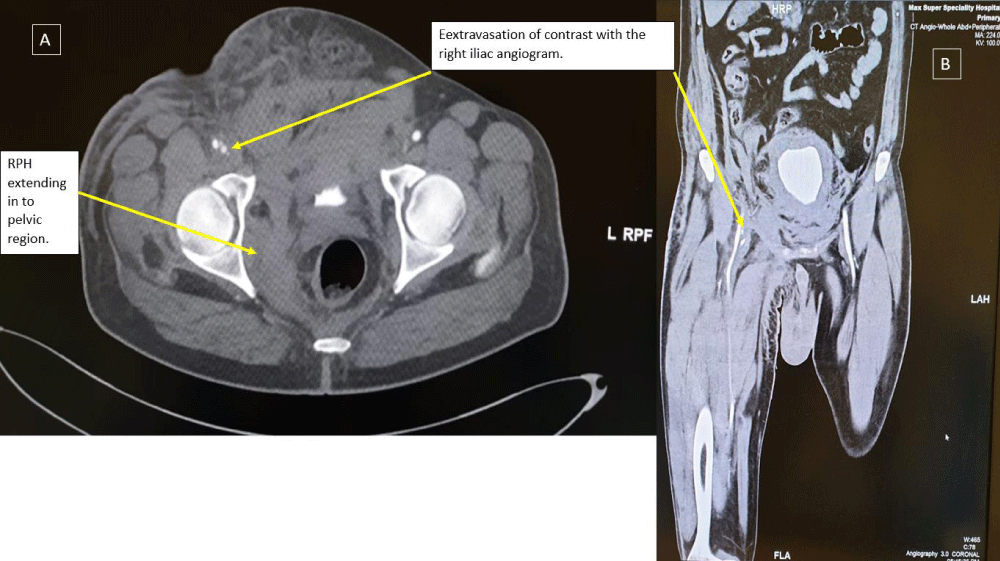
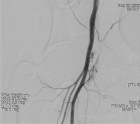
Figure 1:
Contrast computerized tomography of a retroperitoneal hemorrhage are shown in (A) coronal and (B) sagittal views. A large amount of extravascular contrast with the right iliac angiogram location near the middle third of the femoral head, suggestive of active bleeding from mid right common femoral artery into the pelvis.
Read Full Article HTML DOI: 10.29328/journal.jccm.1001119 Cite this Article Read Full Article PDF
More Images
Similar Articles
-
Preclinical stiff heart is a marker of cardiovascular morbimortality in apparently healthy populationCharles Fauvel,Michael Bubenheim,Olivier Raitière,Charlotte Vallet,Nassima Si Belkacem,Fabrice Bauer*. Preclinical stiff heart is a marker of cardiovascular morbimortality in apparently healthy population. . 2019 doi: 10.29328/journal.jccm.1001045; 4: 083-089
-
Left ventricular ejection fraction and contrast induced acute kidney injury in patients undergoing cardiac catheterization: Results of retrospective chart reviewFiras Ajam*,Obiora Maludum,Nene Ugoeke,Hetavi Mahida,Anas Alrefaee,Amy Quinlan DNP,Jennifer Heck-Kanellidis NP,Dawn Calderon DO,Mohammad A Hossain*,Arif Asif. Left ventricular ejection fraction and contrast induced acute kidney injury in patients undergoing cardiac catheterization: Results of retrospective chart review. . 2019 doi: 10.29328/journal.jccm.1001066; 4: 195-198
-
Our experience with single patch repair of complete atrioventricular septal defectsCan Vuran*,Uygar Yoruker,Oguz Omay,Bulent Saritas,Canan Ayabakan,Ozlem Sarisoy,Riza Turkoz. Our experience with single patch repair of complete atrioventricular septal defects. . 2020 doi: 10.29328/journal.jccm.1001095; 5: 105-108
-
Percutaneous treatment of severe retroperitoneal hematoma after percutaneous coronary interventionAgarwal Rajendra Kumar*,Agarwal Rajiv. Percutaneous treatment of severe retroperitoneal hematoma after percutaneous coronary intervention. . 2021 doi: 10.29328/journal.jccm.1001119; 6: 055-058
Recently Viewed
-
Contemplating Catheter Induced Blood Stream Infections and Associated Risk Factors in Diverse Clinical Settings: A Comprehensive ReviewZahra Zahid Piracha, Sadia Mansha, Amna Naeem, Umar Saeed*, Muhammad Nouman Tariq, Azka Sohail, Irfan Ellahi Piracha, Muhammad Shahmeer Fida Rana, Syed Shayan Gilani, Seneen Noor, Elyeen Noor. Contemplating Catheter Induced Blood Stream Infections and Associated Risk Factors in Diverse Clinical Settings: A Comprehensive Review. J Clin Intensive Care Med. 2023: doi: 10.29328/journal.jcicm.1001044; 8: 014-023
-
Sites and Zones of Maximum Reactivity of the most Stable Structure of the Receptor-binding Domain of Wild-type SARS-CoV-2 Spike Protein: A Quantum Density Functional Theory StudyErnesto López-Chávez*, Alberto García-Quiroz, Yesica Antonia Peña-Castañeda, José Antonio Irán Díaz-Góngora, José Alberto Mendoza-Espinosa, J Antonio López-Barrera, Fray de Landa Castillo-Alvarado. Sites and Zones of Maximum Reactivity of the most Stable Structure of the Receptor-binding Domain of Wild-type SARS-CoV-2 Spike Protein: A Quantum Density Functional Theory Study. J Clin Intensive Care Med. 2024: doi: 10.29328/journal.jcicm.1001047; 9: 008-016
-
A Complex Case with a Completely Percutaneous Solution: Treatment of a Severe Calcific Left Main in a Patient with Low-Flow Low-Gradient Aortic StenosisRenatomaria Bianchi*, Giovanni Marco Esposito, Giovanni Ciccarelli, Donato Tartaglione, Paolo Golino. A Complex Case with a Completely Percutaneous Solution: Treatment of a Severe Calcific Left Main in a Patient with Low-Flow Low-Gradient Aortic Stenosis. J Cardiol Cardiovasc Med. 2024: doi: 10.29328/journal.jccm.1001180; 9: 061-066
-
Correlation between the Values of Immature Platelet Fraction and Mean Platelet Volume with the Extent of Coronary Artery Disease in Patients with Non-ST-Segment Elevation Myocardial InfarctionShadab Rauf*, Tarun Kumar, Vijay Kumar, Ranjit Kumar Nath. Correlation between the Values of Immature Platelet Fraction and Mean Platelet Volume with the Extent of Coronary Artery Disease in Patients with Non-ST-Segment Elevation Myocardial Infarction. J Cardiol Cardiovasc Med. 2023: doi: 10.29328/journal.jccm.1001163; 8: 114-121
-
Preventing Coronary Occlusion in an Elderly Severe Aortic Stenosis Patient with Critically Low Coronary Heights – A Case ReportViveka Kumar*. Preventing Coronary Occlusion in an Elderly Severe Aortic Stenosis Patient with Critically Low Coronary Heights – A Case Report. J Cardiol Cardiovasc Med. 2023: doi: 10.29328/journal.jccm.1001165; 8: 130-136
Most Viewed
-
Evaluation of Biostimulants Based on Recovered Protein Hydrolysates from Animal By-products as Plant Growth EnhancersH Pérez-Aguilar*, M Lacruz-Asaro, F Arán-Ais. Evaluation of Biostimulants Based on Recovered Protein Hydrolysates from Animal By-products as Plant Growth Enhancers. J Plant Sci Phytopathol. 2023 doi: 10.29328/journal.jpsp.1001104; 7: 042-047
-
Feasibility study of magnetic sensing for detecting single-neuron action potentialsDenis Tonini,Kai Wu,Renata Saha,Jian-Ping Wang*. Feasibility study of magnetic sensing for detecting single-neuron action potentials. Ann Biomed Sci Eng. 2022 doi: 10.29328/journal.abse.1001018; 6: 019-029
-
Physical activity can change the physiological and psychological circumstances during COVID-19 pandemic: A narrative reviewKhashayar Maroufi*. Physical activity can change the physiological and psychological circumstances during COVID-19 pandemic: A narrative review. J Sports Med Ther. 2021 doi: 10.29328/journal.jsmt.1001051; 6: 001-007
-
Pediatric Dysgerminoma: Unveiling a Rare Ovarian TumorFaten Limaiem*, Khalil Saffar, Ahmed Halouani. Pediatric Dysgerminoma: Unveiling a Rare Ovarian Tumor. Arch Case Rep. 2024 doi: 10.29328/journal.acr.1001087; 8: 010-013
-
Prospective Coronavirus Liver Effects: Available KnowledgeAvishek Mandal*. Prospective Coronavirus Liver Effects: Available Knowledge. Ann Clin Gastroenterol Hepatol. 2023 doi: 10.29328/journal.acgh.1001039; 7: 001-010

HSPI: We're glad you're here. Please click "create a new Query" if you are a new visitor to our website and need further information from us.
If you are already a member of our network and need to keep track of any developments regarding a question you have already submitted, click "take me to my Query."


