Figure 8
Transcatheter Aortic Valve Implantation in Two High-Risk Patients with Low Coronary Ostial Heights Using the Novel Balloon-Expandable Myval Valve
Raja Ramesh N*, Ramesh Daggubati and Ramesh Babu P
Published: 01 August, 2023 | Volume 8 - Issue 2 | Pages: 089-099

Figure 8:
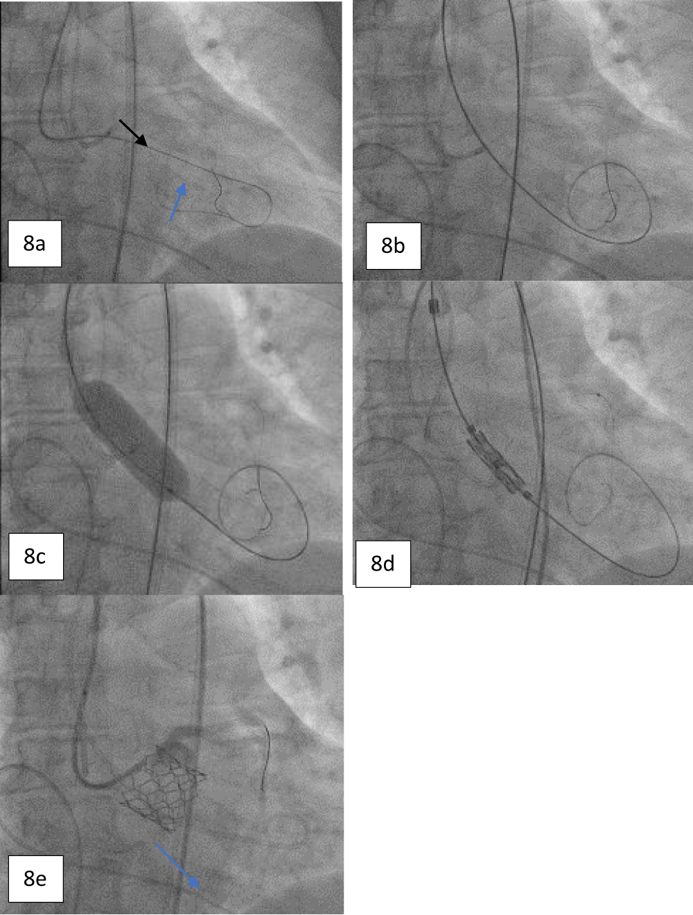
Figure 8: a. Angiogram showing the protection of the LMCA using a guide extension catheter and a supportive coronary guidewire (black arrow) with cannulation; crossing the diseased aortic valve using an AL-1 guiding catheter and a standard straight-tip wire (blue arrow). b. Guidewire exchange using a Safari stiff wire advanced across the valve. c. Pre-dilatation performed with a 20 x 40 mm Mammoth over-the-wire balloon. d. Positioning of 21.5 mm balloon-expandable Myval THV. e. Angiogram obtained post deployment showing the patent LMCA (blue arrow).
Read Full Article HTML DOI: 10.29328/journal.jccm.1001159 Cite this Article Read Full Article PDF
More Images
Similar Articles
-
Early Outcomes of a Next-Generation Balloon-Expandable Transcatheter Heart Valve - The Myval System: A Single-Center Experience From SerbiaDarko Boljevic*, Milovan Bojic, Mihajlo Farkic, Dragan Sagic, Sasa Hinic, Dragan Topic, Milan Dobric, Jovana Lakcevic, Marko Nikolic, Stefan Veljkovic, Matija Furtula, Jelena Kljajevic, Aleksandra Nikolic. Early Outcomes of a Next-Generation Balloon-Expandable Transcatheter Heart Valve - The Myval System: A Single-Center Experience From Serbia. . 2023 doi: 10.29328/journal.jccm.1001156; 8: 072-080
-
Transcatheter Aortic Valve Implantation in Two High-Risk Patients with Low Coronary Ostial Heights Using the Novel Balloon-Expandable Myval ValveRaja Ramesh N*, Ramesh Daggubati, Ramesh Babu P. Transcatheter Aortic Valve Implantation in Two High-Risk Patients with Low Coronary Ostial Heights Using the Novel Balloon-Expandable Myval Valve. . 2023 doi: 10.29328/journal.jccm.1001159; 8: 089-099
-
Preventing Coronary Occlusion in an Elderly Severe Aortic Stenosis Patient with Critically Low Coronary Heights – A Case ReportViveka Kumar*. Preventing Coronary Occlusion in an Elderly Severe Aortic Stenosis Patient with Critically Low Coronary Heights – A Case Report. . 2023 doi: 10.29328/journal.jccm.1001165; 8: 130-136
-
A Complex Case with a Completely Percutaneous Solution: Treatment of a Severe Calcific Left Main in a Patient with Low-Flow Low-Gradient Aortic StenosisRenatomaria Bianchi*, Giovanni Marco Esposito, Giovanni Ciccarelli, Donato Tartaglione, Paolo Golino. A Complex Case with a Completely Percutaneous Solution: Treatment of a Severe Calcific Left Main in a Patient with Low-Flow Low-Gradient Aortic Stenosis. . 2024 doi: 10.29328/journal.jccm.1001180; 9: 061-066
Recently Viewed
-
Navigating Neurodegenerative Disorders: A Comprehensive Review of Current and Emerging Therapies for Neurodegenerative DisordersShashikant Kharat*, Sanjana Mali*, Gayatri Korade, Rakhi Gaykar. Navigating Neurodegenerative Disorders: A Comprehensive Review of Current and Emerging Therapies for Neurodegenerative Disorders. J Neurosci Neurol Disord. 2024: doi: 10.29328/journal.jnnd.1001095; 8: 033-046
-
Statistical Mathematical Analysis of COVID-19 at World LevelMarín-Machuca Olegario*, Carlos Enrique Chinchay-Barragán, Moro-Pisco José Francisco, Vargas-Ayala Jessica Blanca, Machuca-Mines José Ambrosio, María del Pilar Rojas-Rueda, Zambrano-Cabanillas Abel Walter. Statistical Mathematical Analysis of COVID-19 at World Level. Int J Phys Res Appl. 2024: doi: 10.29328/journal.ijpra.1001082; 7: 040-047
-
Electronic and Thermo-Dynamical Properties of Rare Earth RE2X3 (X=O, S) Compounds: A Chemical Bond TheoryPooja Yadav, DS Yadav*, DV Singh. Electronic and Thermo-Dynamical Properties of Rare Earth RE2X3 (X=O, S) Compounds: A Chemical Bond Theory. Int J Phys Res Appl. 2024: doi: 10.29328/journal.ijpra.1001083; 7: 048-052
-
Quantum System Dynamics: Harnessing Constructive Resonance for Technological Advancements, Universal Matter Creation and Exploring the Paradigm of Resonance-induced GravitySanjay Bhushan*. Quantum System Dynamics: Harnessing Constructive Resonance for Technological Advancements, Universal Matter Creation and Exploring the Paradigm of Resonance-induced Gravity. Int J Phys Res Appl. 2024: doi: 10.29328/journal.ijpra.1001084; 7: 053-058
-
Failure-oriented-accelerated-testing (FOAT) and its role in assuring electronics reliability: reviewE Suhir*. Failure-oriented-accelerated-testing (FOAT) and its role in assuring electronics reliability: review. Int J Phys Res Appl. 2023: doi: 10.29328/journal.ijpra.1001048; 6: 001-018
Most Viewed
-
Evaluation of Biostimulants Based on Recovered Protein Hydrolysates from Animal By-products as Plant Growth EnhancersH Pérez-Aguilar*, M Lacruz-Asaro, F Arán-Ais. Evaluation of Biostimulants Based on Recovered Protein Hydrolysates from Animal By-products as Plant Growth Enhancers. J Plant Sci Phytopathol. 2023 doi: 10.29328/journal.jpsp.1001104; 7: 042-047
-
Feasibility study of magnetic sensing for detecting single-neuron action potentialsDenis Tonini,Kai Wu,Renata Saha,Jian-Ping Wang*. Feasibility study of magnetic sensing for detecting single-neuron action potentials. Ann Biomed Sci Eng. 2022 doi: 10.29328/journal.abse.1001018; 6: 019-029
-
Physical activity can change the physiological and psychological circumstances during COVID-19 pandemic: A narrative reviewKhashayar Maroufi*. Physical activity can change the physiological and psychological circumstances during COVID-19 pandemic: A narrative review. J Sports Med Ther. 2021 doi: 10.29328/journal.jsmt.1001051; 6: 001-007
-
Pediatric Dysgerminoma: Unveiling a Rare Ovarian TumorFaten Limaiem*, Khalil Saffar, Ahmed Halouani. Pediatric Dysgerminoma: Unveiling a Rare Ovarian Tumor. Arch Case Rep. 2024 doi: 10.29328/journal.acr.1001087; 8: 010-013
-
Prospective Coronavirus Liver Effects: Available KnowledgeAvishek Mandal*. Prospective Coronavirus Liver Effects: Available Knowledge. Ann Clin Gastroenterol Hepatol. 2023 doi: 10.29328/journal.acgh.1001039; 7: 001-010

HSPI: We're glad you're here. Please click "create a new Query" if you are a new visitor to our website and need further information from us.
If you are already a member of our network and need to keep track of any developments regarding a question you have already submitted, click "take me to my Query."


