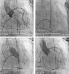
Figure 1
Preventing Coronary Occlusion in an Elderly Severe Aortic Stenosis Patient with Critically Low Coronary Heights – A Case Report
Viveka Kumar*
Published: 19 October, 2023 | Volume 8 - Issue 3 | Pages: 130-136
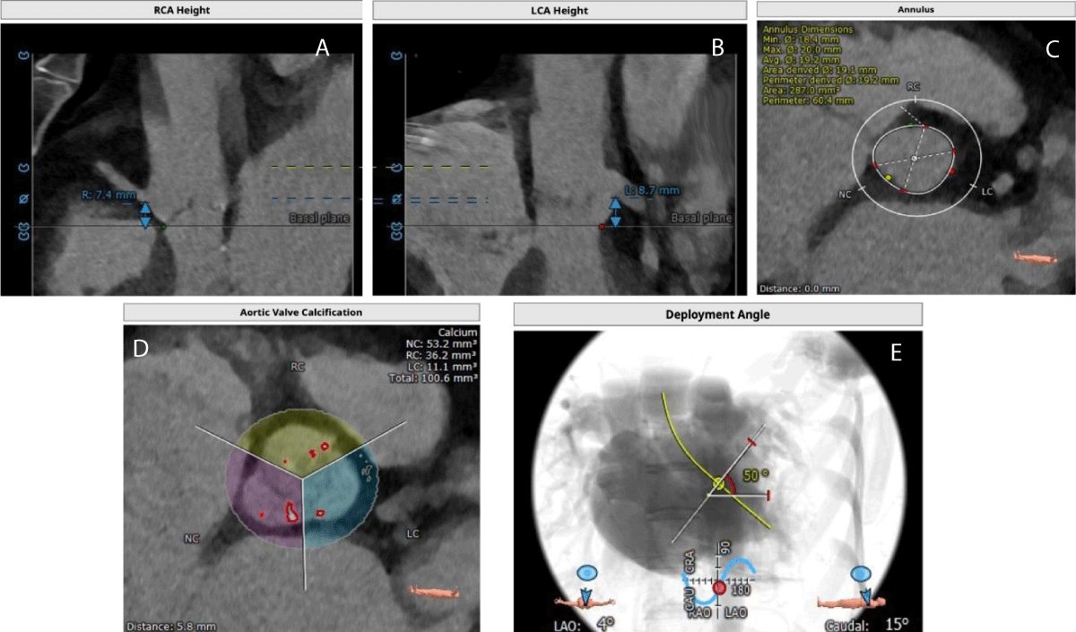
Figure 1:
Figure 1: Computed Tomography images shows- A) Right coronary artery height, B) Left coronary artery height, C) Aortic annulus area, D) Aortic valve calcification and E) Deployment angles.
Read Full Article HTML DOI: 10.29328/journal.jccm.1001165 Cite this Article Read Full Article PDF
More Images
Similar Articles
-
Early Outcomes of a Next-Generation Balloon-Expandable Transcatheter Heart Valve - The Myval System: A Single-Center Experience From SerbiaDarko Boljevic*, Milovan Bojic, Mihajlo Farkic, Dragan Sagic, Sasa Hinic, Dragan Topic, Milan Dobric, Jovana Lakcevic, Marko Nikolic, Stefan Veljkovic, Matija Furtula, Jelena Kljajevic, Aleksandra Nikolic. Early Outcomes of a Next-Generation Balloon-Expandable Transcatheter Heart Valve - The Myval System: A Single-Center Experience From Serbia. . 2023 doi: 10.29328/journal.jccm.1001156; 8: 072-080
-
Transcatheter Aortic Valve Implantation in Two High-Risk Patients with Low Coronary Ostial Heights Using the Novel Balloon-Expandable Myval ValveRaja Ramesh N*, Ramesh Daggubati, Ramesh Babu P. Transcatheter Aortic Valve Implantation in Two High-Risk Patients with Low Coronary Ostial Heights Using the Novel Balloon-Expandable Myval Valve. . 2023 doi: 10.29328/journal.jccm.1001159; 8: 089-099
-
Preventing Coronary Occlusion in an Elderly Severe Aortic Stenosis Patient with Critically Low Coronary Heights – A Case ReportViveka Kumar*. Preventing Coronary Occlusion in an Elderly Severe Aortic Stenosis Patient with Critically Low Coronary Heights – A Case Report. . 2023 doi: 10.29328/journal.jccm.1001165; 8: 130-136
Recently Viewed
-
Diffuse Pediatric-Type High-Grade Glioma H3-/IDH-wildtype with MYCN Deletion and Constitutional Mismatch Repair Deficiency: Case PresentationDarya Sitovskaya*, Mikhail Krapivin, Tatyana Sokolova, Yulia Zabrodskaya. Diffuse Pediatric-Type High-Grade Glioma H3-/IDH-wildtype with MYCN Deletion and Constitutional Mismatch Repair Deficiency: Case Presentation. Arch Case Rep. 2023: doi: 10.29328/journal.acr.1001079; 7: 053-057
-
Fetal Ductal Constriction due to Maternal Intake of MetamizoleRosa Bermejo, Pérez de Heredia Naiara, Faz Cartagena, Francisco Sanchez-Ferrer, Francisco Quereda*. Fetal Ductal Constriction due to Maternal Intake of Metamizole. Arch Case Rep. 2023: doi: 10.29328/journal.acr.1001077; 7: 046-00
-
Morular Metaplasia of the Endometrium: A Case Report and Literature Review: Care Pathways based on Molecular BiologyDhanushka SK Kotigala, Tolu O Adedipe*. Morular Metaplasia of the Endometrium: A Case Report and Literature Review: Care Pathways based on Molecular Biology. Clin J Obstet Gynecol. 2024: doi: 10.29328/journal.cjog.1001165; 7: 059-062
-
Primary Diffuse Leptomeningeal Melanocytosis: A Rare and Challenging DiagnosisStefano Machado*, Diogo Fernandes dos Santos, Andrea De Martino Luppi, Vynícius Vieira Guimarães, Ana Cristina Araújo Lemos da Silva. Primary Diffuse Leptomeningeal Melanocytosis: A Rare and Challenging Diagnosis. J Neurosci Neurol Disord. 2024: doi: 10.29328/journal.jnnd.1001096; 8: 047-049
-
Designing a Community Health Worker (CHW) Certificate Training that Centers Marginalized Youth’s Health and WellnessRuby Mendenhall*, Tramayne Butler-DeLong, Meggan J Lee, Kiara Langford. Designing a Community Health Worker (CHW) Certificate Training that Centers Marginalized Youth’s Health and Wellness. J Community Med Health Solut. 2024: doi: 10.29328/journal.jcmhs.1001047; 5: 052-056
Most Viewed
-
Evaluation of Biostimulants Based on Recovered Protein Hydrolysates from Animal By-products as Plant Growth EnhancersH Pérez-Aguilar*, M Lacruz-Asaro, F Arán-Ais. Evaluation of Biostimulants Based on Recovered Protein Hydrolysates from Animal By-products as Plant Growth Enhancers. J Plant Sci Phytopathol. 2023 doi: 10.29328/journal.jpsp.1001104; 7: 042-047
-
Feasibility study of magnetic sensing for detecting single-neuron action potentialsDenis Tonini,Kai Wu,Renata Saha,Jian-Ping Wang*. Feasibility study of magnetic sensing for detecting single-neuron action potentials. Ann Biomed Sci Eng. 2022 doi: 10.29328/journal.abse.1001018; 6: 019-029
-
Physical activity can change the physiological and psychological circumstances during COVID-19 pandemic: A narrative reviewKhashayar Maroufi*. Physical activity can change the physiological and psychological circumstances during COVID-19 pandemic: A narrative review. J Sports Med Ther. 2021 doi: 10.29328/journal.jsmt.1001051; 6: 001-007
-
Pediatric Dysgerminoma: Unveiling a Rare Ovarian TumorFaten Limaiem*, Khalil Saffar, Ahmed Halouani. Pediatric Dysgerminoma: Unveiling a Rare Ovarian Tumor. Arch Case Rep. 2024 doi: 10.29328/journal.acr.1001087; 8: 010-013
-
Prospective Coronavirus Liver Effects: Available KnowledgeAvishek Mandal*. Prospective Coronavirus Liver Effects: Available Knowledge. Ann Clin Gastroenterol Hepatol. 2023 doi: 10.29328/journal.acgh.1001039; 7: 001-010

HSPI: We're glad you're here. Please click "create a new Query" if you are a new visitor to our website and need further information from us.
If you are already a member of our network and need to keep track of any developments regarding a question you have already submitted, click "take me to my Query."


