Figure 4
The Fulcrum of the Human Heart (Cardiac fulcrum)
Jorge Carlos Trainini*, Mario Wernicke, Mario Beraudo and Alejandro Trainini
Published: 03 January, 2024 | Volume 9 - Issue 1 | Pages: 001-005
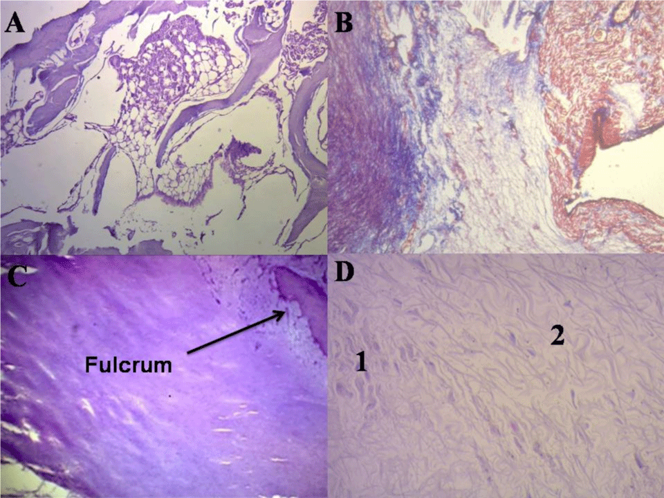
Figure 4:
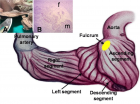
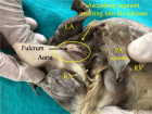
Figure 4: Histology of cardiac fulcrum. A: Mature trabecular bone forming the cardiac fulcrum tissue (bovine heart). Hematoxylin-eosin stain (10x). B: Cardiac fulcrum in a 23-week gestation human fetus showing prechondroid bluish areas in a myxoid stroma. Masson’s Trichrome Technique (15x). C: Central area of the fulcrum formed by chondroid tissue in a10-year-old human heart. Hematoxylin eosin- technique (15x). D: Festooned colagenous bands integrating the fibrotendinous matrix of the fulcrum (adult human heart). Hematoxylin-eosin technique (15x). 1: Remnants of atrophic cardiomyocytes. 2: Fibrotendinous matrix.
Read Full Article HTML DOI: 10.29328/journal.jccm.1001171 Cite this Article Read Full Article PDF
More Images
Similar Articles
-
Thrombolysis, the only Optimally Rapid Reperfusion TreatmentVictor Gurewich*. Thrombolysis, the only Optimally Rapid Reperfusion Treatment. . 2017 doi: 10.29328/journal.jccm.1001010; 2: 029-034
-
A new heart: portraying the physiologic anatomo-functional reconstruction in ischemic cardiomyopathyMarco Cirillo*,Marco Campana,Anna Bressanelli,Giovanni Troise. A new heart: portraying the physiologic anatomo-functional reconstruction in ischemic cardiomyopathy. . 2017 doi: 10.29328/journal.jccm.1001016; 2: 063-067
-
Endogenous sensitizer of beta-adrenergic receptors (ESBAR) and its analogs (review)Victor Tsirkin*,Alexander Nozdrachev,Elena Sizova,Tatyana Polezhaeva,Svetlana Khlybova,Marina Morozova,Andrew Trukhin,Julia Korotaeva,Grigory Khodyrev. Endogenous sensitizer of beta-adrenergic receptors (ESBAR) and its analogs (review). . 2018 doi: 10.29328/journal.jccm.1001028; 3: 064-078
-
Influence of Histidine on the contractility and adrenaline inotropic effect in the experiments with myocardium of right ventricular of Non pregnant and Pregnant RatsVictor Tsirkin*,Alexander Nozdrachev,Julia Korotaeva,Grigorij Khodyrev. Influence of Histidine on the contractility and adrenaline inotropic effect in the experiments with myocardium of right ventricular of Non pregnant and Pregnant Rats . . 2018 doi: 10.29328/journal.jccm.1001030; 3: 084-103
-
The complex interplay in the regulation of cardiac pathophysiologic functionalities by protein kinases and phosphatasesChrysanthus Chukwuma Sr*. The complex interplay in the regulation of cardiac pathophysiologic functionalities by protein kinases and phosphatases. . 2021 doi: 10.29328/journal.jccm.1001118; 6: 048-054
-
The Role of Advanced Imaging in Paediatric Cardiology: Basic Principles and IndicationsMaria Kavga*, Tristan Ramcharan, Kyriaki Papadopoulou-Legbelou. The Role of Advanced Imaging in Paediatric Cardiology: Basic Principles and Indications. . 2023 doi: 10.29328/journal.jccm.1001155; 8: 065-071
-
Rats with Postinfarction Heart Failure: Effects of Propranolol Therapy on Intracellular Calcium Regulation and Left Ventricular FunctionHari Prasad Sonwani*. Rats with Postinfarction Heart Failure: Effects of Propranolol Therapy on Intracellular Calcium Regulation and Left Ventricular Function. . 2023 doi: 10.29328/journal.jccm.1001169; 8: 158-163
-
The Fulcrum of the Human Heart (Cardiac fulcrum)Jorge Carlos Trainini*, Mario Wernicke, Mario Beraudo, Alejandro Trainini. The Fulcrum of the Human Heart (Cardiac fulcrum). . 2024 doi: 10.29328/journal.jccm.1001171; 9: 001-005
Recently Viewed
-
The Comparison of Brachial Artery Parameters between the Clinical Cuff, Pneumatic Controlled Air Band (KAATSU), and Elastic Band during Blood Flow Restriction at the same Perceived TightnessAlexandra Passos Gaspar*, De Matos LDNJ, Amorim S, De Oliveira RS, Fernandes RV, Laurentino G. The Comparison of Brachial Artery Parameters between the Clinical Cuff, Pneumatic Controlled Air Band (KAATSU), and Elastic Band during Blood Flow Restriction at the same Perceived Tightness. J Sports Med Ther. 2024: doi: 10.29328/journal.jsmt.1001076; 9: 015-021
-
Refractory priapism associated with anti-psychotics. Report of a case for risperidoneRamos Luces Odionnys*, Fermín Miriangel and Perdomo Yalisca. Refractory priapism associated with anti-psychotics. Report of a case for risperidone. Arch Case Rep. 2023: doi: 10.29328/journal.acr.1001070; 7: 020-022
-
Incidence of hepatitis B and hepatitis C in Pediatric ward in 2ed March teaching hospital, Sebha: South of LibyaIdress H Attitalla*,Shaban R Bagar,Marei A Altayar,Abdlmanam Fakron,Hosam B Bahnosy. Incidence of hepatitis B and hepatitis C in Pediatric ward in 2ed March teaching hospital, Sebha: South of Libya. Int J Clin Microbiol Biochem Technol. 2021: doi: 10.29328/journal.ijcmbt.1001022; 4: 028-031
-
An uncommon gastrointestinal bleeding in a patient with portal vein thrombosis: a case report and literature reviewDorsa Alijanzadeh, Erfan Arabpour*, Mohammadamin Abdi and Mohammad Abdehagh*. An uncommon gastrointestinal bleeding in a patient with portal vein thrombosis: a case report and literature review. Arch Case Rep. 2023: doi: 10.29328/journal.acr.1001069; 7: 015-019
-
Osteopoikilosis: a rare case with interesting imagingNejadhosseinian Mohammad, Hadighi Pouya, Aghaghazvini Leila, Mozaffari Mohammad Ali, Babagoli Mazyar*, Faezi Seyedeh Tahereh. Osteopoikilosis: a rare case with interesting imaging. Arch Case Rep. 2023: doi: 10.29328/journal.acr.1001068; 7: 012-014
Most Viewed
-
Evaluation of Biostimulants Based on Recovered Protein Hydrolysates from Animal By-products as Plant Growth EnhancersH Pérez-Aguilar*, M Lacruz-Asaro, F Arán-Ais. Evaluation of Biostimulants Based on Recovered Protein Hydrolysates from Animal By-products as Plant Growth Enhancers. J Plant Sci Phytopathol. 2023 doi: 10.29328/journal.jpsp.1001104; 7: 042-047
-
Feasibility study of magnetic sensing for detecting single-neuron action potentialsDenis Tonini,Kai Wu,Renata Saha,Jian-Ping Wang*. Feasibility study of magnetic sensing for detecting single-neuron action potentials. Ann Biomed Sci Eng. 2022 doi: 10.29328/journal.abse.1001018; 6: 019-029
-
Physical activity can change the physiological and psychological circumstances during COVID-19 pandemic: A narrative reviewKhashayar Maroufi*. Physical activity can change the physiological and psychological circumstances during COVID-19 pandemic: A narrative review. J Sports Med Ther. 2021 doi: 10.29328/journal.jsmt.1001051; 6: 001-007
-
Pediatric Dysgerminoma: Unveiling a Rare Ovarian TumorFaten Limaiem*, Khalil Saffar, Ahmed Halouani. Pediatric Dysgerminoma: Unveiling a Rare Ovarian Tumor. Arch Case Rep. 2024 doi: 10.29328/journal.acr.1001087; 8: 010-013
-
Prospective Coronavirus Liver Effects: Available KnowledgeAvishek Mandal*. Prospective Coronavirus Liver Effects: Available Knowledge. Ann Clin Gastroenterol Hepatol. 2023 doi: 10.29328/journal.acgh.1001039; 7: 001-010

HSPI: We're glad you're here. Please click "create a new Query" if you are a new visitor to our website and need further information from us.
If you are already a member of our network and need to keep track of any developments regarding a question you have already submitted, click "take me to my Query."


